Types of Brain AVM: Size, Location, and Risk Factors
The human brain is an intricate network of blood vessels, neurons, and tissues working together to maintain life. One rare but serious condition affecting this system is an Arteriovenous Malformation (AVM). This abnormal connection between arteries and veins disrupts normal blood flow and oxygen circulation, often posing significant health risks. Understanding the types of brain AVM, their classification based on size, location, and associated risk factors, is vital for early detection and treatment.
For patients in Maharashtra, consulting an experienced brain specialist in Pune is crucial for accurate diagnosis, advanced treatment planning, and long-term management of brain AVMs.
What is a Brain AVM?
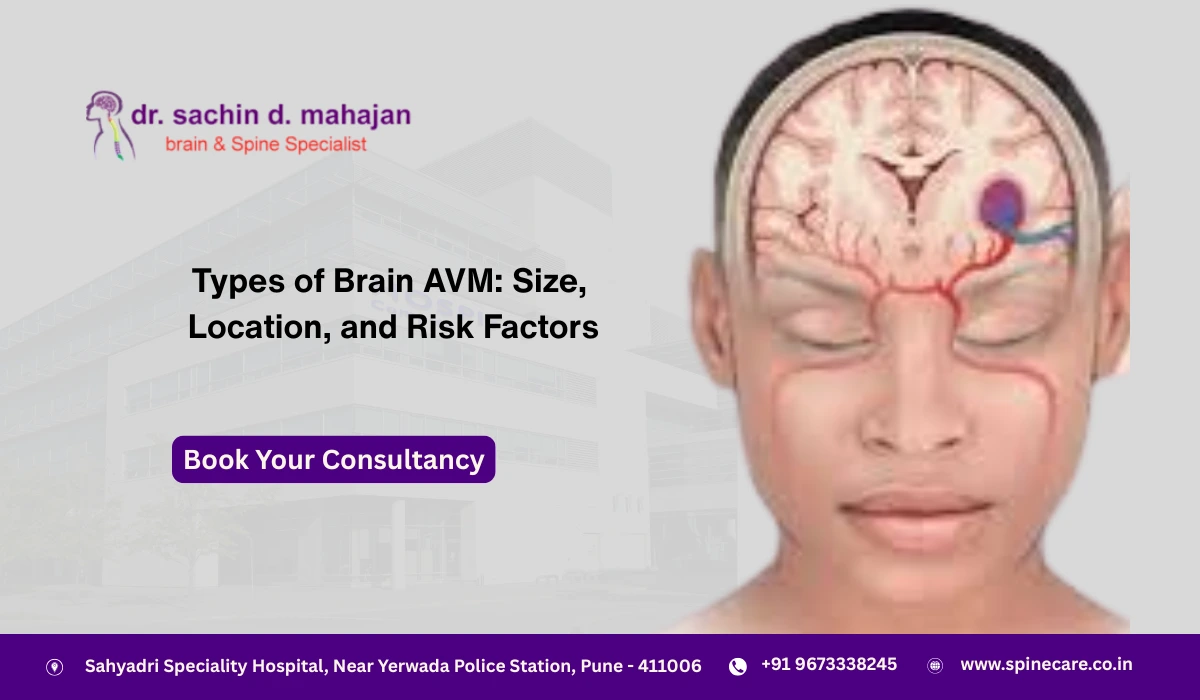
A brain AVM is a tangle of abnormal blood vessels that connect arteries and veins, bypassing the capillary system. This direct connection increases pressure and risk of rupture, potentially leading to bleeding in the brain (hemorrhage), seizures, headaches, or neurological issues.
AVMs can occur anywhere in the brain or spinal cord, and their impact depends on factors such as size, location, and associated risk factors.
Classification of Brain AVMs
Medical experts classify AVMs to determine the best treatment approach. The most common classification methods are based on size, location, and risk factors.
1. Classification by Size
AVMs are usually divided into three size categories:
- Small AVMs (less than 3 cm in diameter):
- Easier to treat surgically or with radiosurgery.
- Lower risk of surgical complications but may still cause bleeding.
- Medium AVMs (3–6 cm in diameter):
- Pose higher treatment challenges.
- Require advanced techniques, often combining surgery, embolization, or radiosurgery.
- Large AVMs (greater than 6 cm in diameter):
- Complex and riskier to treat.
- Often located in deep or sensitive areas of the brain.
- High risk of hemorrhage and neurological deficits.
2. Classification by Location
The brain is divided into functional regions, and the location of an AVM greatly influences symptoms, risks, and treatment decisions.
- Superficial (Cortical) AVMs:
- Found on the surface of the brain.
- Often associated with seizures or headaches.
- Deep AVMs:
- Located in critical brain structures such as the basal ganglia, brainstem, or thalamus.
- More challenging to treat due to delicate surroundings.
- Infratentorial AVMs (Cerebellum/Brainstem):
- Can affect balance, coordination, and vital functions.
- Tend to have higher rupture risks.
- Supratentorial AVMs (Cerebral Hemispheres):
- May cause motor weakness, sensory loss, or visual disturbances.
3. Classification by Risk Factors
Doctors assess risks using factors like:
- Age of the patient: Younger patients have longer exposure risk.
- History of hemorrhage: Previous bleeding increases future rupture chances.
- AVM size and location: Larger and deeper AVMs are more dangerous.
- Associated aneurysms: Presence of aneurysms raises rupture risks.
- Venous drainage pattern: AVMs with deep venous drainage are more hazardous.
Symptoms of Brain AVM
Not all AVMs cause symptoms, but when they do, the signs can be life-threatening. Common symptoms include:
- Severe headaches or migraines
- Seizures
- Weakness or numbness in limbs
- Vision problems
- Difficulty with speech or understanding
- Loss of coordination or balance
If you experience sudden, severe neurological symptoms, immediate medical attention from a brain specialist in Pune is essential.
Diagnosis of Brain AVM
To confirm an AVM, specialists use advanced imaging tests:
- MRI and MRA (Magnetic Resonance Imaging/Angiography)
- CT Scan
- Cerebral Angiogram (Gold Standard Test)
These tests help identify the size, location, and structure of the AVM for tailored treatment.
Treatment Options for Brain AVM
The treatment approach depends on whether the AVM has ruptured and the patient’s overall health. Some AVMs may be monitored, while others require urgent intervention.
- Microsurgical Resection
- Surgical removal of the AVM.
- Often preferred for small, accessible AVMs.
- Endovascular Embolization
- A minimally invasive procedure using catheters to block abnormal blood vessels.
- Often used in combination with surgery or radiosurgery.
- Stereotactic Radiosurgery (Gamma Knife/CyberKnife):
- Focused radiation shrinks the AVM over time.
- Suitable for small to medium AVMs in difficult locations.
- Conservative Management:
- Monitoring for asymptomatic or very high-risk AVMs where surgery is not feasible.
Risk Factors and Complications
Brain AVMs can cause severe complications if untreated:
- Hemorrhage (Bleeding in the brain): The most serious complication.
- Stroke: Reduced blood flow may damage brain tissue.
- Seizures: Due to abnormal electrical activity.
- Neurological Deficits: Weakness, speech problems, or paralysis.
Patients should not ignore headaches or seizure episodes. Seeking timely care ensures better treatment outcomes.
Importance of Specialized Neurological Care in Pune
When dealing with complex conditions like AVM, specialized care is non-negotiable. Pune is home to advanced neurosurgical centers offering state-of-the-art diagnostic and treatment facilities.
An experienced brain specialist in Pune provides:
- Accurate AVM diagnosis with advanced imaging.
- Individualized treatment planning.
- Access to minimally invasive surgical procedures.
- Long-term rehabilitation and follow-up care.
For conditions such as AVMs, aneurysms, or even severe facial pain disorders like Trigeminal Neuralgia Treatment In Pune, patients benefit from expert neurosurgical evaluation and advanced treatment techniques.
Lifestyle and Recovery After AVM Treatment
- Follow-Up Scans: Regular MRI/CT scans to monitor treated AVM.
- Medication: To manage seizures or prevent bleeding.
- Rehabilitation: Physiotherapy, speech therapy, or occupational therapy for recovery.
- Lifestyle Adjustments: Stress reduction, balanced diet, and avoiding smoking/alcohol.
Conclusion
Brain AVMs are rare but serious conditions requiring careful evaluation and treatment. Understanding their types—based on size, location, and risk factors—helps patients and families make informed decisions.
If you or a loved one experiences neurological symptoms, seeking timely consultation with a brain Tumor specialist in Pune is the first step toward effective care. Expert neurosurgeons also provide advanced solutions like Trigeminal Neuralgia Treatment In Pune, ensuring comprehensive brain and spine care under one roof.
CTA (Call to Action):
Looking for advanced care for brain AVM or other neurological conditions? Consult the leading brain specialist in Pune at SpineCare and explore modern treatment options for safer, faster recovery.